AllFocusTM
EDF
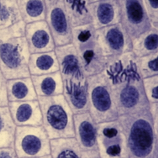
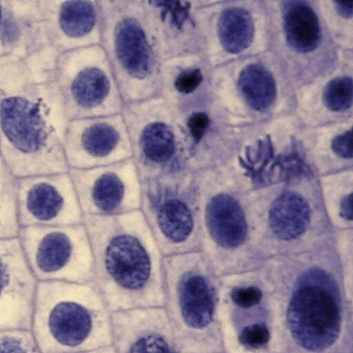
Image Gallery - Section of Onion Root Tip
Thick section of an
Onion
Root Tip
from a slide made
by Alexis Scientific
www.alsci.com
shows interphase and mitotic cells. Images were acquired on Olympus BX51 with UPlanFl 40X
0.75NA objective in transmitted light.
Condenser was set
at 0.75NA. Focus was motorized by the
Focus
Drive with Integrated Controller. Camera -
Canon Digital Rebel
positioned in primary image plane. Control software -
ZStackTool and
Canon Remote Capture. Z-stack
of 9 images was acquired with step size of 1 micron and processed with
AllFocus V0.97.
Top left image is the
Z-stack animation, top right image is the standard Merged Image produced by AllFocus.
In this case the
merge method was linear combination of images in the stack with weights
determined by focus function. Bottom two images illustrate the ability of
AllFocus to assign higher weight to the beginning or to the end of the stack.
The feature of interest is the two interphase cells positioned vertically under
each other in the center of the frame. Along the depth of the stack a
mitotic cell underneath these two is revealed. The standard
Merged Image shows the overall best focus image. The bottom left image has
preference for the beginning of the stack and the bottom right image has
preference for the end of the stack. The weight changes linearly along the
stack and is defined by a single number.